John Kendrew,
Max Perutz
chemistry

|
Protein crystallography
The first proteins whose structures were determined by X-ray crystallography were myoglobin, the protein that stores oxygen in animal muscle tissues, and hemoglobin, the protein that transports oxygen in the bloodstream. John Kendrew and Max Perutz studied hemoglobin from sheep but were not able to make much progress. Obtaining myoglobin from a whale, which formed crystals that were large enough, Kendrew and Perutz compared X-ray diffraction patterns of various samples infused with different metals.
The structure of myoglobin
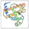
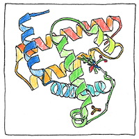
A myoglobin is a polypeptide with eight connected right-handed alpha-helices. Hydrophobic amino acid groups are packed in myoglobin’s interior and hydrophilic amino acid groups are exposed on its surface. One heme prosthetic group is packed into a hydrophobic cleft. Each heme group has one iron atom that binds to an oxygen atom.
The structure of hemoglobin
A hemoglobin contains four polypeptide clusters alpha, beta, gamma, delta, each containing the same kind of heme prosthetic group found in myoglobin.
Red-blooded
The red color of blood doesn’t come from the iron in it, but from the porphyrin to which the iron is bound. Nobody’s skin is naturally red and everybody’s blood is red, and the average person has average passions. Being a great singer or artist requires hard work and long study; being an American, only being born here.


It would be difficult to overestimate the difficulty of this work. It took Dorothy Hodgkin 35 years to fuss out the structure of insulin. Kendrew and Perutz had to devise innovative tricks and techniques to get from a frozen block of protein sufficient information about these complex structures.
See also in The book of science:
Readings in wikipedia:
Other readings: